| |

Professor and
Nuclear Medicine Specialist Jean Verreault of Sherbrooke Hospital, says major advances in PET Imaging will have
a profound
impact in the field of Oncology for the future. |
|
NUCLEAR MEDICINE
SPECIALIST
PROFESSOR JEAN VERREAULT DISCUSSES THE ADVANCES IN CLINICAL PET IMAGING
AND ITS POTENTIAL IMPACT ON ARAB HEALTH
Professor
Jean Verreault, M.D., F.R.C.P., specialist in nuclear medicine at
Sherbrooke Hospital in Quebec, Canada was invited by Philips Medical
Systems to address delegates on 'Advances in Clinical PET Imaging' attending the Middle East Imaging &
Diagnostic Conference held as part of Arab Health '2003 (26-29 January, Dubai,
UAE). Professor Verreault sat down in a special interview
to
discuss those advances in clinical PET imaging - its relationship to
technology, its costs and benefits, and its future for improving
healthcare in the Arab World with ArabMedicare.com
CEO Mahatma Davis.
ArabMedicare.com: Explain
briefly how diagnosis and
recognition of disease using diagnostic imaging has undergone significant
change and development during the past few years?
Verreault: The noninvasive evaluation of myocardial viability
in the 80s and more recently the
explosion of PET (Positron Emission Tomography) imaging in oncology has
had a profound impact in the detection and management of many
diseases. These have had a positive impact on the industry. PET will
probably be the major change occurring for the next 5 to 10 years.
Up to now, there have been a lot of efforts in developing high-resolution anatomy
pictures but we are more and more moving to functional imaging with PET
which is now called "molecular imaging" referring directly to PET. In PET imaging, advancements in new
tracers or specific tracers have been developed for detecting malignant
tumors mainly because of the use of 18 F. The main one used in
oncology is 18 FDG (F-18 fluorodeoxyglucose is used as a marker for glucose metabolism, which, in
turn is an indicator of cellular activity). This has given physicians the ability
to better manage patients with cancer and to determine best therapy
approaches that take into account the precise staging of the disease.
ArabMedicare.com: What dramatic changes do you
envision for the next 5-10 years?
Verreault: The development of PET imaging as the major tool
in the field of oncology. PET is a powerful diagnostic tool that in many
cases renders answers that no other imaging tests can provide. The
combination of high-resolution anatomy pictures from CT and functional
imaging with PET will become the main tool
for physicians in diagnosis and treatment specially in the areas of oncology,
cardiology, and neurology. PET imaging can pinpoint the areas of disease
activity, as well as evaluate response to treatment, thus, providing crucial
information for disease management.
ArabMedicare.com: What areas
of PET imaging research do you consider should be given more importance?
Verreault: In imaging in general, there has been a lot of effort
pushed on high-resolution anatomy. But more and more, we are moving
towards "molecular imaging" and its direct relationship to PET. I do believe that this trend is definitely here to stay.
Furthermore, in the area of research, there has been significant developments
in devices. But more new work has to come in developing specific tracers like those that will be able to image slow
growing
tumors that can be mist with FDG imaging.
ArabMedicare.com: Due to its
current limited availability, what are some of the challenges that you
foresee for using PET in the Arab World, as compared to your experience
with other health systems?
Verreault: It would have been hard to think of having PET
scanning 10-12 years ago in every big hospital because each time you
installed a camera you would have need a cyclotron beside it due to the
short live of isotopes previously used. This is not the
case anymore with FDG since you can put a cyclotron in a designated
location and distribute by network FDG taking about 2 hours turnaround time. So
you can now have a few hospitals operating their own dedicated PET system
and buying their FDG from the same source. This has lowered the
overall cost and made PET more accessible, practical, and affordable. Also, the
improvement and the speed of the camera have lowered the cost of PET scans
during the
last few years so that more exams can be done during the same period of
time.
However, for the region, strategically, the
first step would be to install these cyclotron centers to be shared by
several hospitals. After this you have to be sure that you have
competent people to operate the systems. At the moment, it is not
clear if there will be enough people to meet the demands of all of the
cyclotrons that will be installed in the region. The region will
need people with experience in radiopharmaceuticals as well as trained in how to
run the systems.
This will be one of the main problems that will need to be addressed by the
industry during the early stages of planning and building a PET imaging
infrastructure for the region.
After that the next step would be to
install dedicated PET systems. I hope that the hospitals will not try to
upgrade existing systems to coincide with PET imaging. This would be
an error. PET imaging is a very powerful diagnostic tool, but you
have to do it right and with good tools.
|
| |
ArabMedicare.com: Can you give
us examples of some of the successes with PET imaging?
Verreault: The main one is that we are able to
identify a disease that is far more advanced than previously
known. This way we can inform the patient that it would not be useful to subject
him to a surgery given the fact his cancer
is too far advance. It is sad to say, but at least it is the
truth and the patient knows what to expect and how to plan for the
remainder of his life. The other more positive one is that we are able to confirm
to the patient that he is disease free after chemotherapy.
ArabMedicare.com: How
do you view the use of PET in combination with other devices such as
CT scanners?
Verreault: Adding the PET to the CT scanner will most
definitely increase the cost. It will also provide more
integrated reports. However, I am not sure it is a must at the
moment. In Sherbrooke Hospital, we use two different systems,
which were purchased over time. If we had to make a similar choice
today, we would request for one system to be PET/CT.
ArabMedicare.com: What are the most
frequently covered PET indications?
Verreault: The main use of PET in oncology is the
evaluation of lung cancer. It represents about 30-33% of our
activity in Canada. The second one is the evaluation of lymphoma
especially for controlling the therapy after chemotherapy
treatment. We will do a PET scan to assure that the
treatment is complete. Other frequent PET indications include colon
and breast cancer diseases.
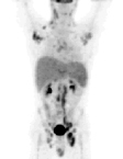
(PET Imaging in extensive Lymphoma)
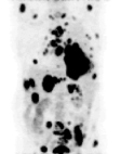
(PET Imaging in
metastatic
Lung Cancer)
ArabMedicare.com: What
are some of the cost factors involved with using PET imaging, and
how can Arab hospitals prepare their health systems for adopting
PET technology?
Verreault: It depends on how the governments
will develop their policies. It will require a comprehensive
structure in which cost will indeed be a factor. In the
public sector, patient care is the most important factor.
For the private practice, patient care is equally important but cost must
be justified in order to sustain the facility and the ability to
delivery quality care.
PET has been quite successful in
demonstrating cost reductions in private hospitals by reducing the
number of unnecessary surgical procedures and by earlier detection
of disease followed by more preventive measures to be implemented.
In most instances, early detection and appropriate treatment
selection can have a major impact on the cost as well as the
outcome of the treatment. For example, a single PET scan can give information about
cancer activity in the entire body. This can be a crucial advantage
in many instances since additional tumors focus and a more
extensive spread of disease are sometimes discovered. The
replacement of multiple tests is also a great convenience for patients
and physicians, as the extent of disease is established quickly
and with greater confidence. This should also be taken into
account when talking about costs.
The
increase and adoption for the use of PET technology in the Arab World will
provide new options for local patients to seek diagnosis,
treatment, and related medical services at home
versus traveling abroad.
ArabMedicare.com: What are some
of the main ethics issues that confront the use of PET?
Verreault: As with every technology, each society has to make its
choice. Should we put more money on prevention, on
diagnostic, on therapeutics? All of these aspects have to be compared
together. But, with PET, I am sure that if we can diagnose
cancer more precisely and sooner, we will get a better
staging of the disease and the therapeutic aspect will benefit
from it. Prevention will also be helped since we will be
able to make some
diagnostics sooner in the evolution of the disease.
One could almost consider it unethical
in 2003 not to use PET given the advantages of
detection and diagnosis that it can offer to the
quality of care for the patient.
|
|
|