|
Researchers use medical
ultrasound to examine coral
reproduction for the first
time
(DUBAI)
-
Coral Spawning International
(CSI), in partnership with
Canon Medical Systems UK
and Canon EMEA, has
conducted what it describes
as the world’s first
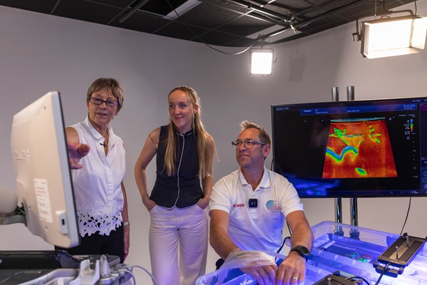
ultrasound scan of living
coral to identify developing
eggs. The experiment used
Canon’s Aplio i700
ultrasound system and could
provide researchers with a
non-invasive way to study
coral reproduction.
Six species of large polyp
stony (LPS) coral, each with
different skeletal
structures, were scanned
while fully submerged in
water. The research aimed to
investigate whether
ultrasound could identify
developing eggs without the
need to cut into coral
specimens, as is required by
some traditional methods of
examining their internal
reproductive structures.
Using high- and
ultra-high-frequency probes,
researchers were able to
image internal coral
structures in detail. The
findings suggest that
ultrasound could help
identify developing eggs and
improve researchers’ ability
to predict spawning periods,
potentially informing coral
conservation, breeding, and
restoration programs.
To verify the scan
results, a specimen of
Trachyphyllia geoffroyi was
fragmented and examined.
Macro photography of the
coral’s internal structures
was then compared with the
ultrasound images. This
confirmed that the
structures identified in the
scans were eggs, providing
initial evidence that
ultrasound can be used to
detect developing eggs in
living coral.
Dr Jamie Craggs,
Co-Founder of Coral Spawning
International, said:
“Corals still hold so many
secrets, particularly when
it comes to the complex
processes that drive their
reproduction. Being able to
look inside a living coral
and see these processes
unfolding in such remarkable
detail is an extraordinary
moment. This experiment has
given us a new perspective
on these animals and, as we
continue monitoring them
through spawning, we hope to
build an increasingly
detailed picture of how
different coral species
reproduce.”
Sally Davey, Clinical
Product Specialist at Canon
Medical Systems UK, said:
“This experiment showcased
just how powerful ultrasound
technology can be. Moreover,
the experiment was
successful and opens
opportunities in fields such
as minimally invasive
surgery and diagnosis of
dermal conditions.”
Peter Bragg,
Sustainability and
Government Affairs Director
at Canon EMEA, added:
“This experiment
demonstrates that imaging
innovation has the power to
show us what was previously
unseen, creating a deeper
understanding of the world
around us. By unlocking this
knowledge, we hold a
fundamental belief that
imaging has the power to
transform the world.”
The six coral species
will continue to be
monitored over six months,
with observations recorded
when they release gametes.
The researchers aim to use
this data to develop a
better understanding of LPS
coral reproductive cycles
and assess whether
ultrasound can help predict
spawning periods more
accurately, supporting
future conservation and
restoration work.
 PRINT
THIS ARTICLE
|